
 11
11


A starry sky can be stunningeven inside a hospital emergency room.But instead of celestial bodies sparkling in the night, doctors in South Korea were gazing at bright brain lesions punctuating a magnetic resonance imaging (MRI) scan.
The resulting pattern, called a "starry sky," meant that their 57-year-old patient had a dangerous form of tuberculosis.
The doctors report the case in this week's issue of the New England Journal of Medicine.The man had previously been treated for the infection in his lungs but came into the hospital's emergency department after two weeks of unexplained headaches, neck pain, and tingling in his right hand.
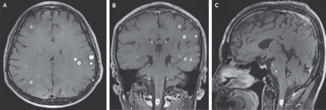
The MRI and Computed-Tomography (CT) scans clearly revealed the problem: rare nodules and lesions, called tuberculomas, speckling his lungs and central nervous system, including both cerebral hemispheres, the basal ganglia deep inside the brain, the cerebellum at the back of the brain, the brain stem, and the upper spinal cord.
Magnetic resonance imaging (MRI) of the head with gadolinium enhancement revealed numerous small, spherical, peripherally enhancing nodules in the cerebral hemispheres (Panels A and B), basal ganglia, cerebellum, and brain stem, as well as in the upper spinal cord with surrounding edema (Panel C).
Credit: NEJM, 2025 The condition, called CNS tuberculoma, is a relatively rare manifestation of tuberculosis, which typically infects the lungs but can invade any part of the body.
It's unclear exactly how tuberculomas form, but evidence suggests that the bacteria that cause tuberculosisMycobacterium tuberculosiscan spread around the body via the blood.M.
tuberculosiscan get past the blood-brain barrier, possibly by hiding inside a type of white blood cell called a macrophage, in a "Trojan horse" mechanism or by breaking through the barrier.Tuberculomas are thought to form when bacteria and macrophages clump together into masses that may contain calcifications or cheese-like dead tissue called caseum.